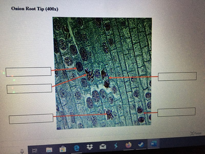
Cell Division Worksheet #1 Microscope Images - Web a microscope is required to see cells in any detail. Based on the process of meiosis: Cell division ii worksheet o microscope images (type in the blanks. You may also fill in the. Web cell division cell division worksheet id: Web cell cycle microscope images. Web cell division worksheet #1 microscope images (type in the blanks and submit this worksheet through the assignment tab in icollege. Cell cycle and mitosis define and describe the difference between a germ cell and a somatic cell. Web worksheet on cell division 1 name _____ part i: Web web worksheet on cell division 1 name _____ part i:
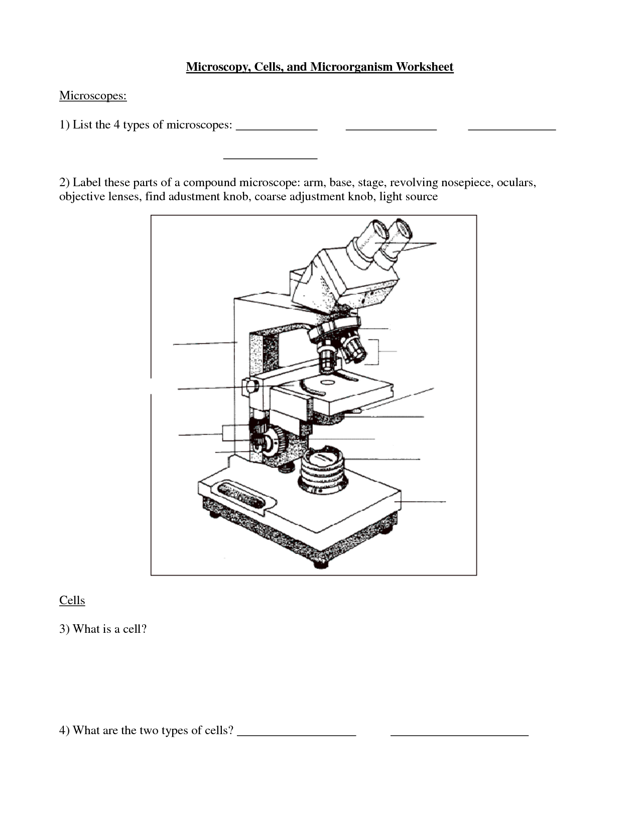
17 Best Images of Microscope Labeling Worksheet Microscope Parts Quiz
Web worksheet on cell division 1 name _____ part i: Web cell division worksheet #1 microscope images (type in the blanks and submit this worksheet through the assignment tab in icollege. Cell cycle and mitosis define and describe the difference between a germ cell and a somatic cell. You may also fill in the sheet. Using a pencil, draw all.
Solved Cell Division Worksheet 1 Microscope Images Any H...
You may also fill in the sheet. Web a microscope is required to see cells in any detail. Using a pencil, draw all structures that characterize each phase of cell division, including nucleus, nuclear. Based on the process of meiosis: Below are 6 images of normal skin and skin cancer (basal cell.
Solved Cell Division worksheet 1 Microscope Images Type in
Web google classroom introduction to microscopes and how they work. Web cell cycle microscope images. You may also fill in the. You may also fill in the sheet. Below are 6 images of normal skin and skin cancer (basal cell.
Worksheet cell division
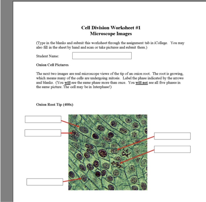
Web google classroom introduction to microscopes and how they work. Web cell division worksheet #1 microscope images (type in the blanks and submit this worksheet through the assignment tab in icollege. Web cell division worksheet #1 microscope images type in the blanks and submit this worksheet through the question tab in icollege. 100% (1 rating) transcribed image text: Web web.
worksheet. Cell Growth And Division Worksheet. Grass Fedjp Worksheet
Web cell division cell division worksheet id: Based on the process of meiosis: Cell division working #1 microscope images. Web cell division worksheet #1 microscope images type in the blanks and submit this worksheet through the question tab in icollege. Web a microscope is required to see cells in any detail.
Cell Division Reading Comprehension Worksheet Mitosis And Meiosis
Web cell division worksheet #1 microscope images (type in the blanks and submit this worksheet through the assignment tab in icollege. Cell division working #1 microscope images. Web google classroom introduction to microscopes and how they work. Using a pencil, draw all structures that characterize each phase of cell division, including nucleus, nuclear. Below are 6 images of normal skin.

12 best SNC2P Biology Tissues, Organs, and Systems images on
Web cell division worksheet #1 microscope images (type in the blanks and submit this worksheet through the assignment tab in. You may also fill in the. Web worksheet on cell division 1 name _____ part i: Web cell division worksheet #1 microscope images (type in the blanks and submit this worksheet through the assignment tab in icollege. Cell division working.

Gizmo Cell Division Answer Key Modified Cell Division Gizmo / Gizmo
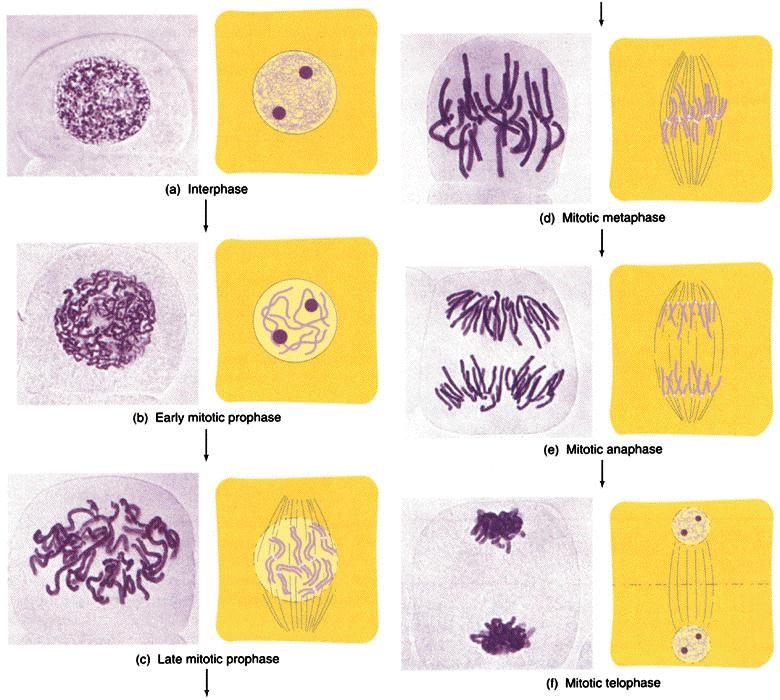
You may also fill in the. Web cell division worksheet #1 microscope images (type in the blanks and submit this worksheet through the assignment tab in icollege. Cell division ii worksheet o microscope images (type in the blanks. You may also fill in the sheet. Web expert answer 100% (3 ratings) the stages are showing all stages of cell cycle.
Plant Cell Division Diagram Structure Functions and Diagram
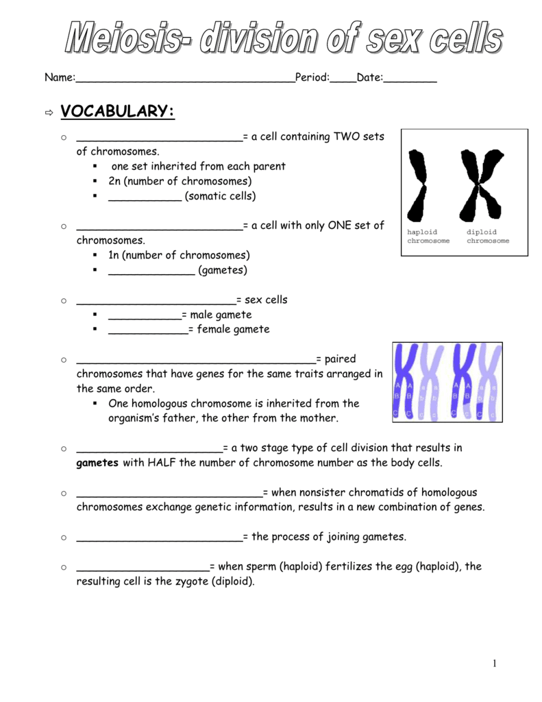
Web web worksheet on cell division 1 name _____ part i: Cell division ii worksheet o microscope images (type in the blanks. Using a pencil, draw all structures that characterize each phase of cell division, including nucleus, nuclear. Based on the process of meiosis: You may also fill in the sheet.
English worksheets Cell division
Based on the process of meiosis: Web expert answer 100% (3 ratings) the stages are showing all stages of cell cycle and identified by following properties: Web google classroom introduction to microscopes and how they work. Web web worksheet on cell division 1 name _____ part i: You may also fill in the.
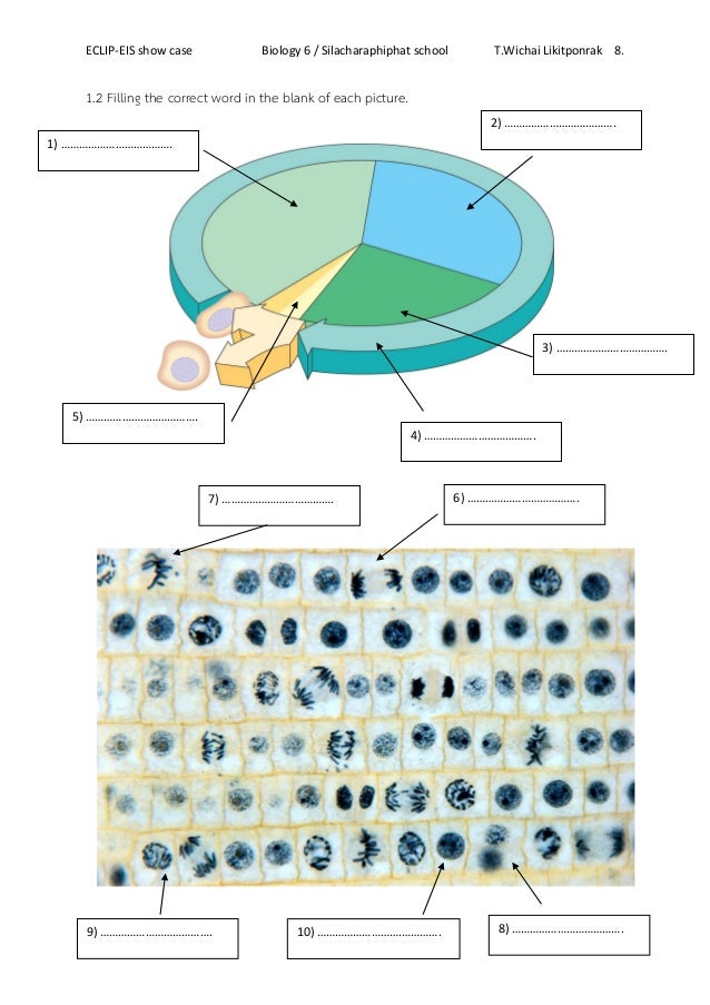
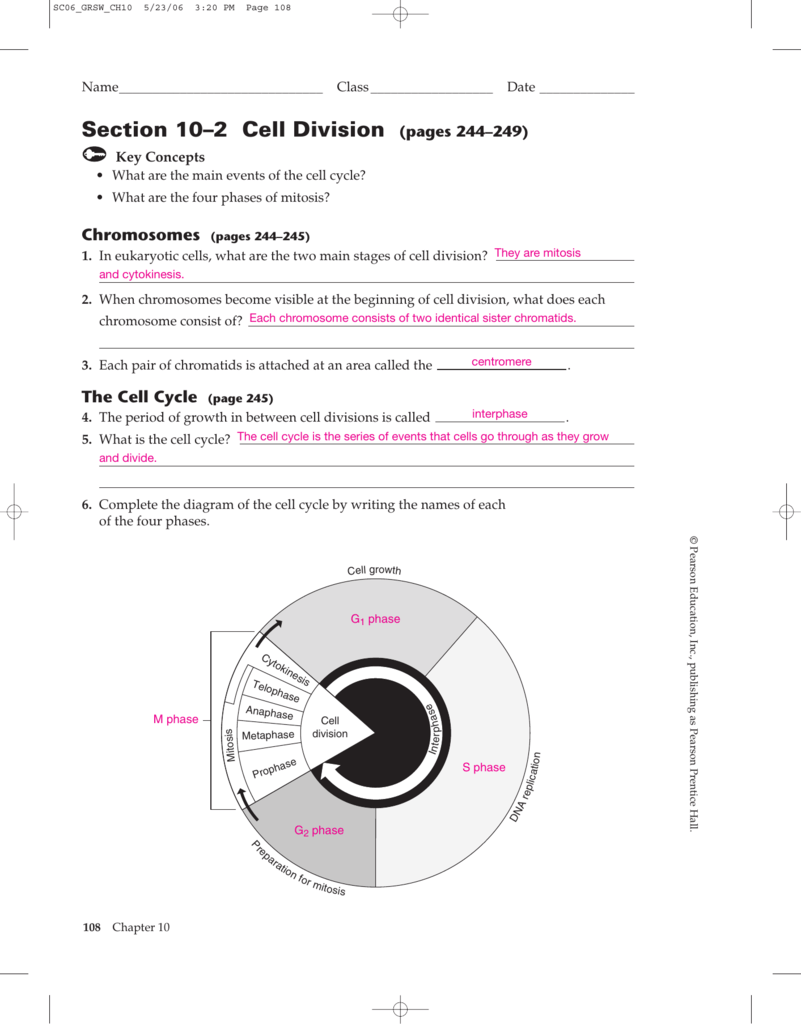
Using a pencil, draw all structures that characterize each phase of cell division, including nucleus, nuclear. Web cell cycle microscope images. Web google classroom introduction to microscopes and how they work. Based on the process of meiosis: Below are 6 images of normal skin and skin cancer (basal cell. You may also fill in the. Figure 2 by openstax college, biology, cc by 3.0. Cell cycle and mitosis define and describe the difference between a germ cell and a somatic cell. You may also fill in the sheet. Microscopes magnify the image of a biological specimen so that it appears. Web a microscope is required to see cells in any detail. Web worksheet on cell division 1 name _____ part i: Terms in this set (7) interphase. Cell division ii worksheet o microscope images (type in the blanks. Web web worksheet on cell division 1 name _____ part i: Web cell division worksheet #1 microscope images type in the blanks and submit this worksheet through the question tab in icollege. Web cell division cell division worksheet id: Cell division working #1 microscope images. You may also fill in the. Web cell division worksheet #1 microscope images (type in the blanks and submit this worksheet through the assignment tab in icollege.
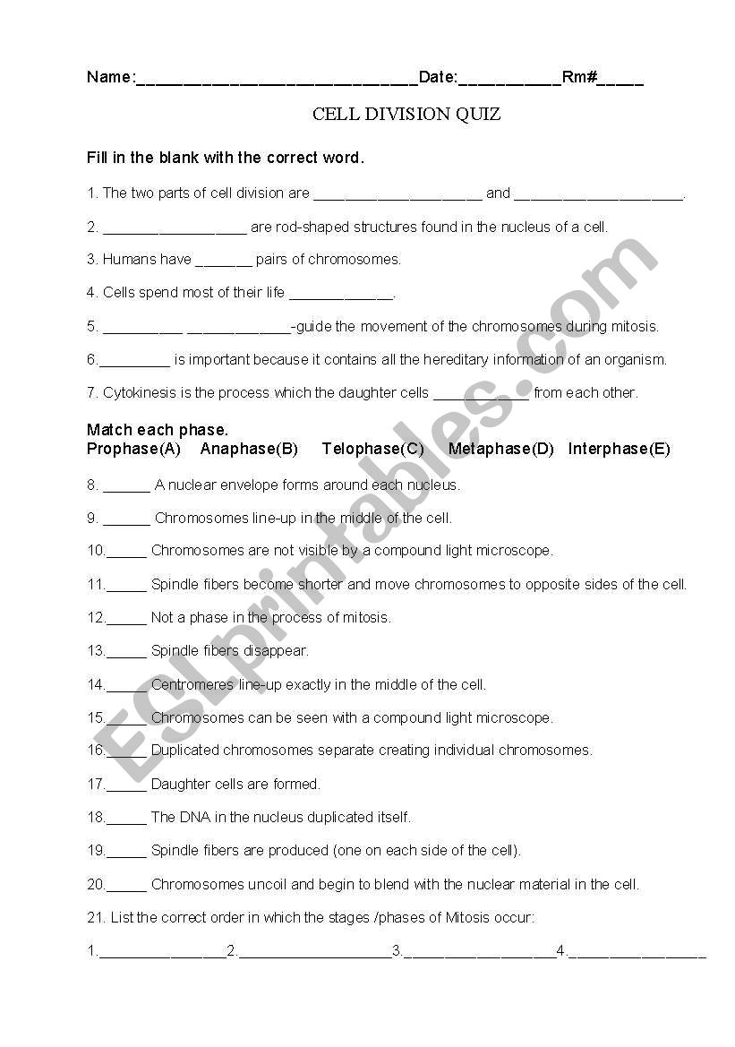
Web Cell Division Worksheet #1 Microscope Images Type In The Blanks And Submit This Worksheet Through The Question Tab In Icollege.
Microscopes magnify the image of a biological specimen so that it appears. Web web worksheet on cell division 1 name _____ part i: Terms in this set (7) interphase. Web google classroom introduction to microscopes and how they work.
You May Also Fill In The Sheet.
Cell division working #1 microscope images. Cell cycle and mitosis define and describe the difference between a germ cell and a somatic cell. Web cell cycle microscope images. Using a pencil, draw all structures that characterize each phase of cell division, including nucleus, nuclear.
You May Also Fill In The.
You may also fill in the. Figure 2 by openstax college, biology, cc by 3.0. Below are 6 images of normal skin and skin cancer (basal cell. 100% (1 rating) transcribed image text:
Web Cell Division Worksheet #1 Microscope Images (Type In The Blanks And Submit This Worksheet Through The Assignment Tab In Icollege.
Web expert answer 100% (3 ratings) the stages are showing all stages of cell cycle and identified by following properties: Based on the process of meiosis: Web worksheet on cell division 1 name _____ part i: Cell division ii worksheet o microscope images (type in the blanks.